WHAT IS A LATERAL ANKLE SPRAIN?
There are three lateral ligaments in the ankle. They are the anterior talofibular ligament, calcaneofibular ligament, and posterior talofibular ligament. A sprain of the lateral ligaments occurs when the ankle is in the open-packed position of plantarflexion and inversion. The anterior talofibular ligament is most commonly sprained, followed by the calcaneofibular ligament, and then the posterior talofibular ligament. A minor tear is classified as a grade I tear, followed by grade II if there is significant tearing, and grade III if the ligament is completely torn. The two main tests that test for a lateral ankle sprain are the anterior drawer test and the inversion talar tilt test. Below is a description of what clinical findings are indicative of a lateral ankle sprain.
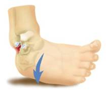 Inversion motion causes ankle sprain.
History
- Pain: Lateral ankle around malleolus and sinus tarsi
- Onset: Acute
- Mechanism: Inversion (supination, plantarflexion, and talar rotation)
- Swelling around lateral joint capsule
- Ecchymosis (discoloration) around lateral malleolus
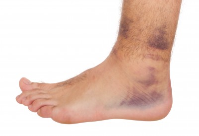 Lateral swelling and ecchymosis.
Palpation
- Pain with palpation of lateral ligaments and sinus tarsi
- Active ROM: pain with plantarflexion and inversion
- Passive ROM: pain with platerflexion, inversion in neutral, inversion with dorsiflexion
